Case Feed
TeslaViewer_344 · EXT-2026-EE544D
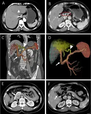
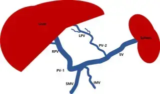
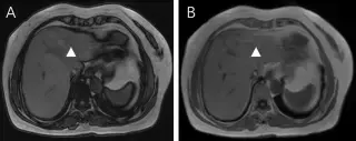
67-year-old woman, asymptomatic, suspected liver space-occupying lesion on routine ultrasound. Normal liver function tests, negative viral hepatitis markers.
GammaFocus_882 · EXT-2026-3B48D0
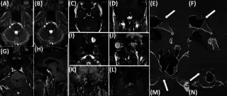
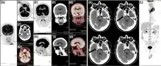
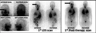
39-year-old male with diplopia and neck swelling. Thyroid FNAC revealed follicular neoplasm.
ParenchymalViewer_870 · EXT-2026-7267D1
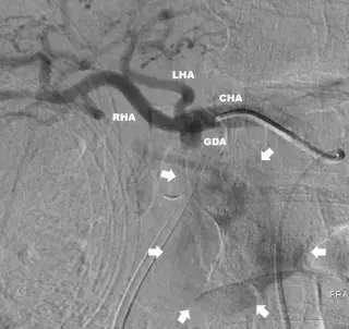
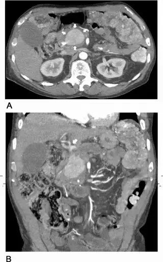
74-year-old male with chronic alcoholic pancreatitis, presenting with progressive epigastric abdominal pain and nausea. Lab studies showed mild transaminitis and hyperbilirubinemia.
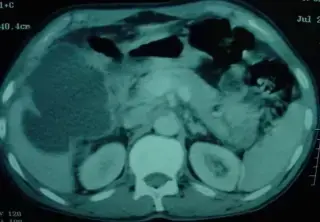
EchoGantry_373 · EXT-2026-0966EE
65-year old man, presented with abdominal pain, nausea and vomiting of 48 hours duration.
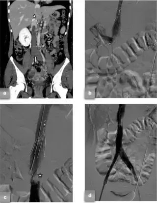
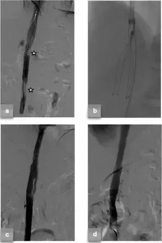
TeslaViewer_344 · EXT-2026-2377F7
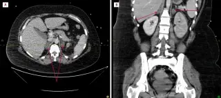
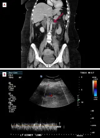
20-year-old female, 10th week of pregnancy, acute abdominal pain. History of congenital left renal atrophy with compensatory right renal hypertrophy, no prior thrombotic events.
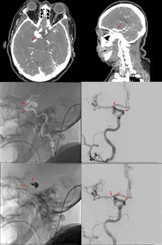
EchoScout_273 · EXT-2026-2CF16F
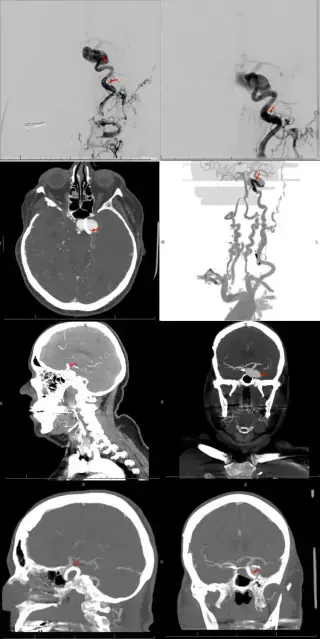
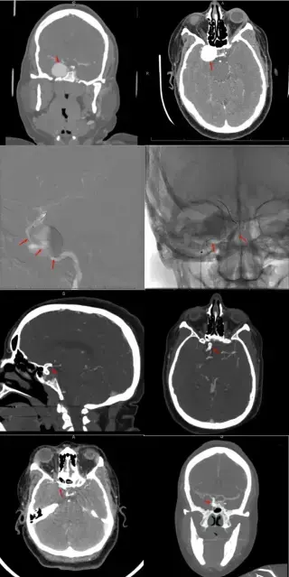
69-year-old patient with left-sided facial and upper/lower extremity hemiparesis. History of hyperlipidemia, hypertension, and 50-pack year smoking.
GammaFocus_882 · EXT-2026-80E5D5
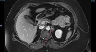
27-year-old female with chest and flank pain, 4 hours after normal vaginal delivery of her first child. Re-presented 17 days later with worsening bilateral pleuritic chest and flank pain.
GantryGantry_464 · EXT-2026-B1FD5C
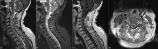
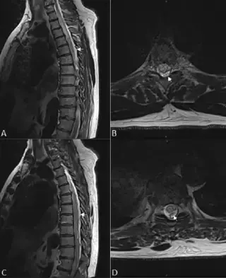
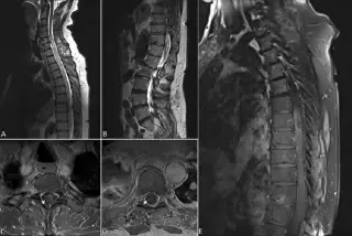
65F with hyperlipidemia, osteoporosis. Progressive right upper extremity weakness, difficulty ambulating, severe neck pain, paresthesias, rapidly growing cervical spinal cord mass.
IsointenseFocus_271 · EXT-2026-8FB9BA
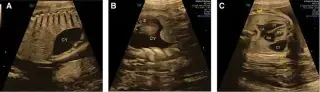
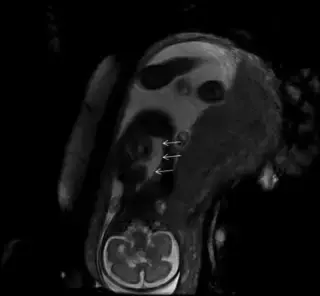
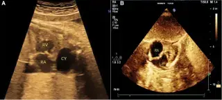
34F, G4P2, 28 weeks pregnant, referred for pericardial effusion diagnosed at 22 5/7 weeks. External ultrasound showed 6.5mm fluid dark area beside right cardiac wall.
QuantumExplorer_803 · EXT-2026-51C5E7
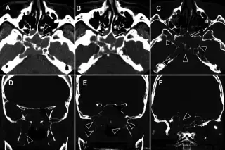
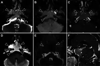
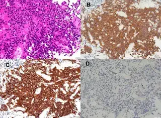
64-year-old woman referred for paranasal sinus tumor evaluation. History of hypertension, bilateral hip replacement, severe mitral regurgitation. Sinus tumor incidentally found on MRI during preoperative exam. No subjective symptoms.
SpectralScan_327 · EXT-2026-61426F
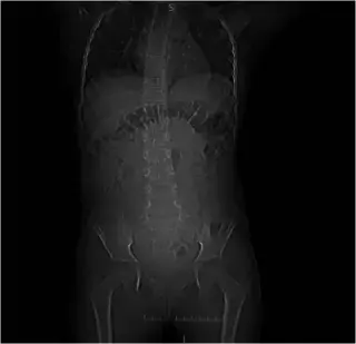
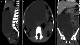
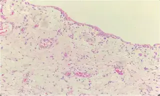
9-year-old girl with 1-week severe colicky abdominal pain, distension, 4 days vomiting (initially undigested food/gastric juice, later bloody). No prior surgery, pre-menarcheal.
TeslaWave_474 · EXT-2026-FB9E35
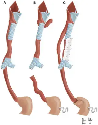
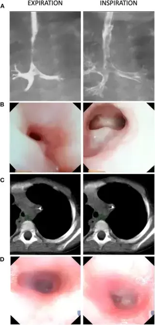
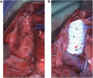
Female neonate, 38 weeks gestational age, 2,770g birth weight. Prenatal ultrasound detected right kidney agenesis and suspected aortic coarctation.
Showing 229–240 of 297 cases