Case Feed
EchoScout_273 · EXT-2026-614695
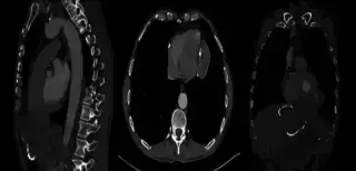
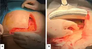
65-year-old male with two months of right upper quadrant pain, left-sided chest pain, and epigastric discomfort. No past medical history. Physical exam showed epigastric dullness. Labs normal.
FocalMatrix_426 · EXT-2026-0489FB
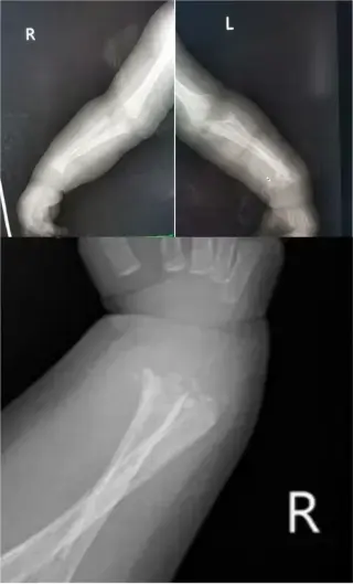
2-month old male infant presented for investigation of bilateral ulna and distal radius fractures. Also had noisy breathing, runny nose, reduced feeding. Mother noticed inability to move upper extremities and crying with passive movement a few days prior.
CoronalVoxel_131 · EXT-2026-9AC9DB
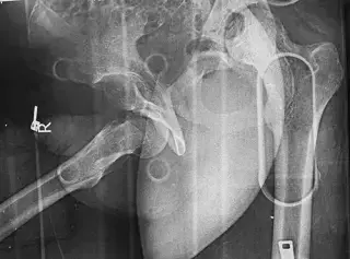
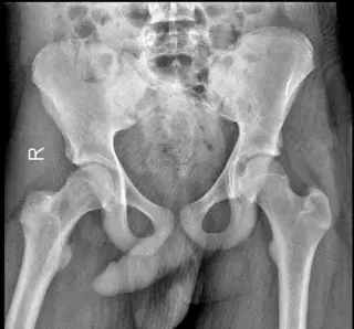
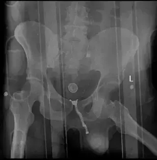
17-year-old male, severe pain after roadside accident, hit by automobile from lateral side, rolled over, could not remember moment exactly.
GantryGantry_464 · EXT-2026-667424
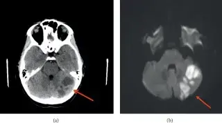
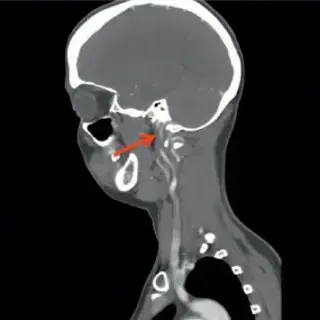
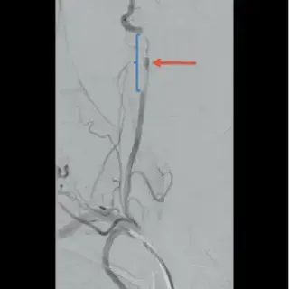
37-year-old female with dyslipidemia, asthma, and fibroids presented with two weeks of bilateral posterior neck pain and headaches, attributed to physical activities and recent roller coaster rides.
HyperSlice_562 · EXT-2026-279D0C
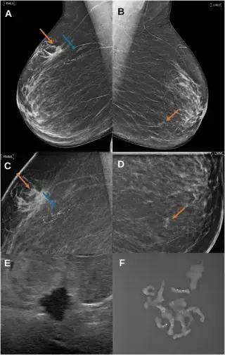
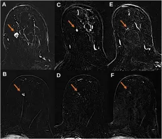
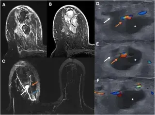
64F, postmenopausal, hormonal substitution, hx surgically treated VIN 3, family hx gynecological cancers, positive breast cancer screening.
CoronalVoxel_131 · EXT-2026-BC5D0C
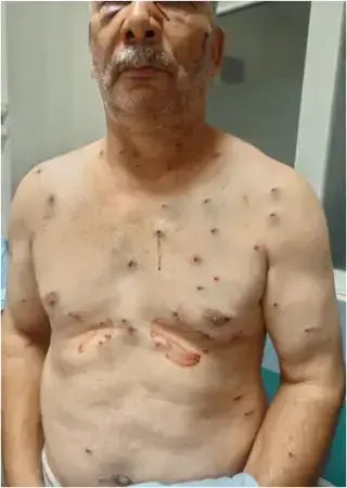
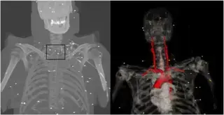
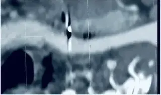
45-year-old male with active smoking history, presented 2 hours after accidental shotgun discharge to the neck. No significant past medical/surgical history.
SpecularPhantom_904 · EXT-2026-D8BDB0
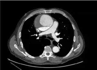
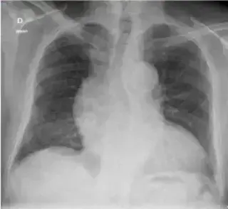
70-year-old man with mild chest pain. History of treated arterial hypertension. Physical exam revealed a diastolic heart murmur. ECG was normal.
QuantumNode_728 · EXT-2026-6C68FC
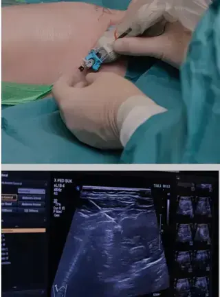
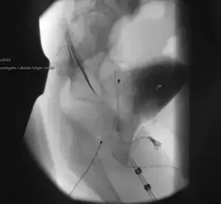
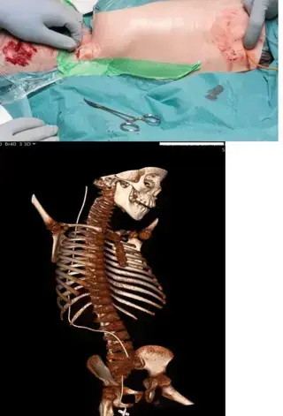
Premature boy (26+2 weeks, 1068g) with history of necrotizing enterocolitis, grade IV intraventricular haemorrhage, and posthemorrhagic hydrocephalus. Underwent two laparotomies.
EchoGantry_373 · EXT-2026-0E0736
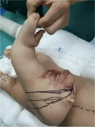
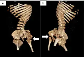
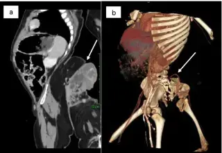
12-month-old boy with a sacrococcygeal mass, detected by prenatal US at 34 weeks gestation. Physical exam revealed a 35x25cm lower limb-like structure.
ParenchymalViewer_870 · EXT-2026-D130AC
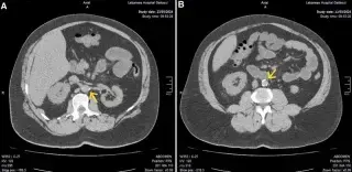
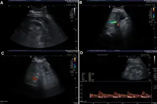
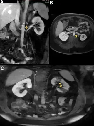
47M with Behcet's disease, hypertension, dyslipidemia, CAD, ED, and gout, presented with severe, continuous left flank pain radiating to the left groin, developing gradually over 3 days.
ParenchymalViewer_870 · EXT-2026-B6939F
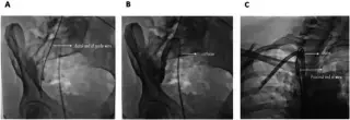
45-year-old female with 13-year history of uncontrolled hypertension and ESRD on hemodialysis, referred for incidental finding of retained guidewire left during venous catheter placement 3 months prior.
FocalScan_107 · EXT-2026-A695A0
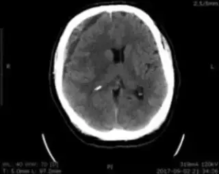
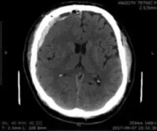
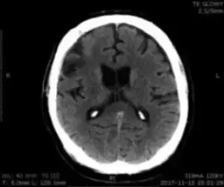
66-year-old male with hypertension suffered head injury in car accident. Six weeks later admitted to neurosurgery. Conscious with full verbal contact on exam.
Showing 241–252 of 297 cases