Case Feed
AxiomNode_187 · EXT-2026-4F21B9
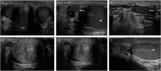
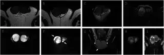
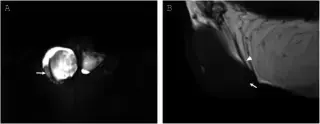
53-year-old man with a firm, painless right scrotal mass, history of prostate cancer treated with radical prostatectomy and lymph node dissection.
HyperdenseBeam_813 · EXT-2026-AACE7F
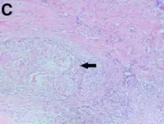
39-year-old lady with a 3-month history of an indurated mass lesion and non-healing ulcer in the medial part of the right lower lid.
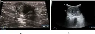
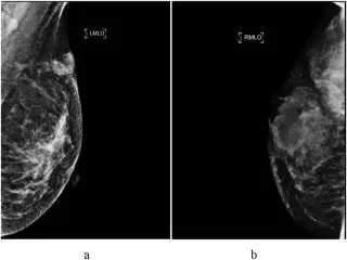
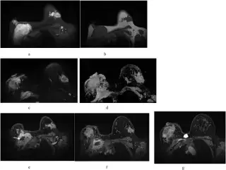
SpecularPhantom_904 · EXT-2026-918D2D
27-year-old woman, presented with a tender right breast mass first palpated 3 years earlier, which gradually enlarged during pregnancy and remained stable after delivery.
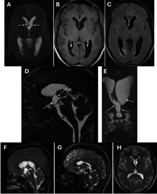

QuantumNode_728 · EXT-2026-3C1A24
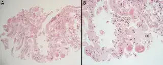
Unknown age, unknown sex. Complaint: Acute obstructive hydrocephalus.
ParenchymalSeeker_124 · EXT-2026-5CAA06
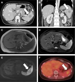
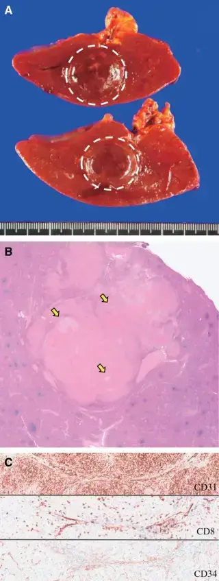
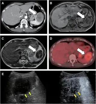
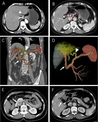
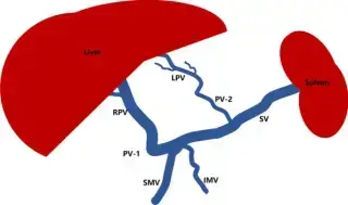
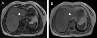
47-year-old female followed-up for gallbladder polyps. Receiving medical treatment for pulmonary arterial hypertension. Referred for detailed examination.
TeslaViewer_344 · EXT-2026-EE544D
67-year-old woman, asymptomatic, suspected liver space-occupying lesion on routine ultrasound. Normal liver function tests, negative viral hepatitis markers.
GantryWindow_627 · EXT-2026-5E3128
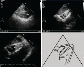
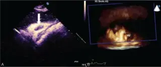
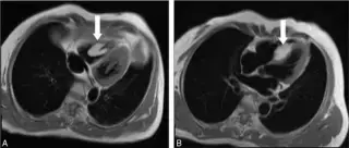
48-year-old woman with 10 years of occasional palpitation on exertion, worsening for 6 months. Found to have a cardiac mass 2 weeks prior. Unremarkable medical/family history, weight loss.
ParenchymalSeeker_124 · EXT-2026-5510BB
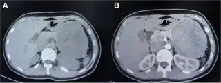
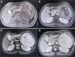
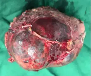
17-year-old girl with AMH for 6 weeks, admitted after ultrasound diagnosis of a pancreatic mass. No pain, urinary symptoms, abdominal distension, fever, jaundice, nausea, vomiting, heartburn, weight loss, or loss of appetite.

EchoGantry_373 · EXT-2026-0E0736
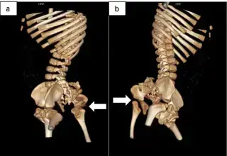
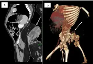
12-month-old boy with a sacrococcygeal mass, detected by prenatal US at 34 weeks gestation. Physical exam revealed a 35x25cm lower limb-like structure.
GammaFocus_882 · EXT-2026-80E5D5
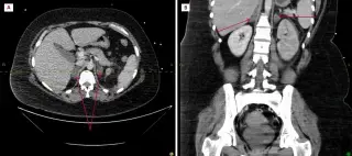
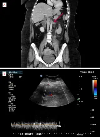
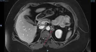
27-year-old female with chest and flank pain, 4 hours after normal vaginal delivery of her first child. Re-presented 17 days later with worsening bilateral pleuritic chest and flank pain.
GantryGantry_464 · EXT-2026-911F62
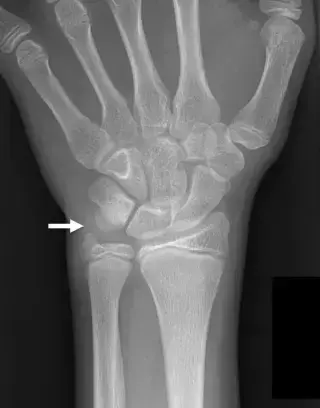
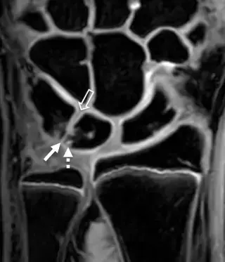
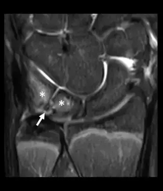
13-year-old boy with left wrist pain with active use. History of left wrist injury 3 months prior in soccer. Pre-existing chronic ulnar-sided wrist pain with repetitive use and limited range of motion.
SpecularPhantom_904 · EXT-2026-D8BDB0
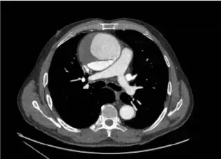
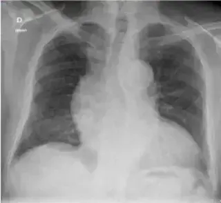
70-year-old man with mild chest pain. History of treated arterial hypertension. Physical exam revealed a diastolic heart murmur. ECG was normal.
Showing 13–24 of 34 cases