Key Images. Click to enlarge

Clinical History
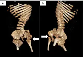
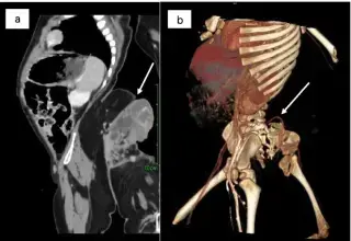
12-month-old boy with a sacrococcygeal mass, detected by prenatal US at 34 weeks gestation. Physical exam revealed a 35x25cm lower limb-like structure.
Write your interpretation
Sign in to submit your read. The original report is revealed only after you submit.