Case Feed
ParenchymalSeeker_124 · EXT-2026-B14AD1
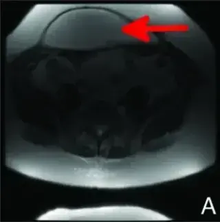
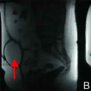
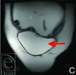
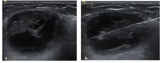
50-year-old woman with Cowden's syndrome and hypothyroidism, post-prophylactic mastectomies and breast reconstruction. First seroma aspiration 2 weeks post-op.
HyperSlice_562 · EXT-2026-84E79A
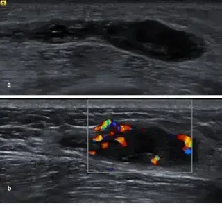
Female with recent trauma history.
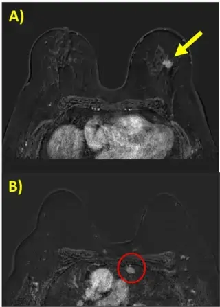
ParenchymalSeeker_124 · EXT-2026-FEFEA3
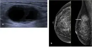
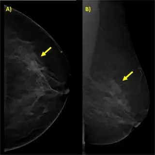
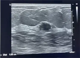
64-year-old woman with a palpable left breast lump. No associated symptoms. No family history of breast cancer. Menopause at 50. Sedentary, on beta-blocker for occasional hypertension.
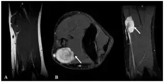
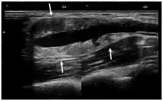
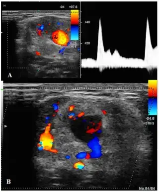
ParenchymalSeeker_124 · EXT-2026-3F3BC9
36-year-old man with a painless, palpable, mobile, soft, pulsating, non-tender mass in the right arm near the cubital fossa, slowly growing for 5 years. No skin changes, pulses present.
TeslaWave_474 · EXT-2026-5B669C
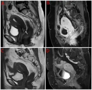
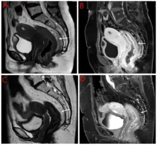
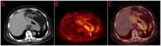
64-year-old postmenopausal woman with 10-year history of hypertension, presenting with urinary urgency, frequent urination, altered stool morphology, abdominal distension, and pain.
ParenchymalFocus_598 · EXT-2026-1138F7
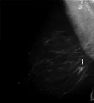
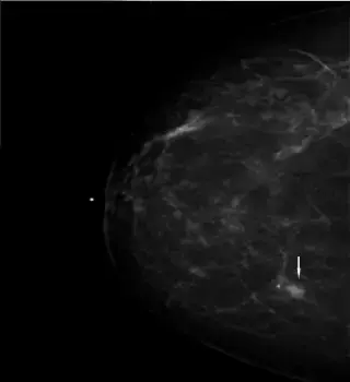
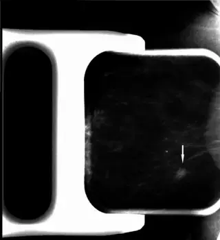
A woman in her seventh decade presented for screening mammography. She had a history of lumpectomy, chemotherapy, and radiation therapy for stage IA ER+/PR+/HER2- IDC and DCIS 16 years prior.
ParenchymalFocus_598 · EXT-2026-F1FD01
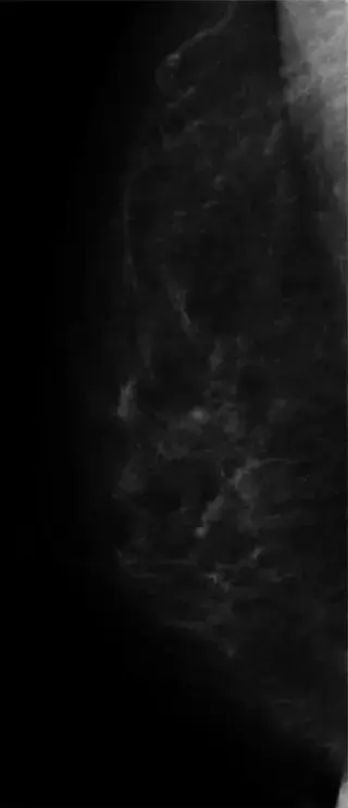
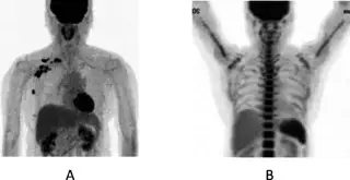
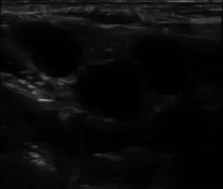
62F with history of left micro invasive breast cancer 15 years prior, treated with breast-conserving therapy, sentinel node biopsy, and 5 years adjuvant tamoxifen. Annual check-ups.
HyperSlice_562 · EXT-2026-0339F7
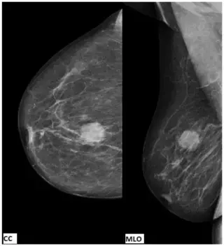
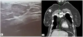
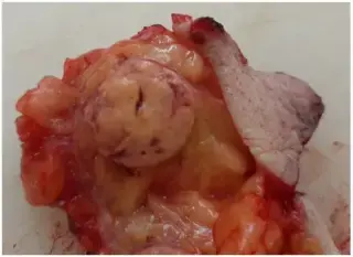
82-year-old female with history of treated renal cell carcinoma, presenting for complete diagnostics due to new appearance/symptoms.
TeslaWave_474 · EXT-2026-46D263
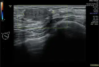
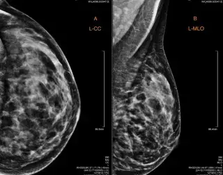
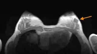
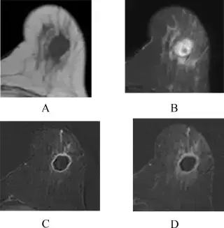
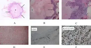
A 32-year-old woman presented with a palpable, well-defined, non-tender, mobile mass in her left breast, located at the four o’clock position, 3 cm from the nipple, with a maximum diameter of 2.1 cm.
HyperSlice_562 · EXT-2026-7E6181
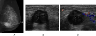
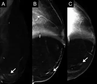
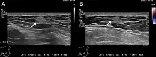
54-year-old woman with a left breast mass.
SpecularPhantom_904 · EXT-2026-F46431
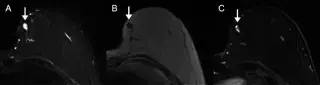
64-year-old female with history of left breast DCIS s/p mastectomy with reconstruction 10 years prior, no recurrence. Presented for routine screening mammography, recalled for new mass.
ParenchymalFocus_598 · EXT-2026-978CA4
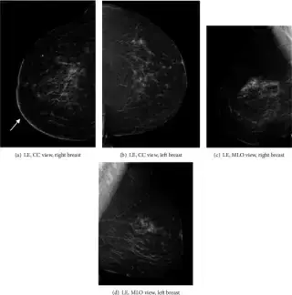
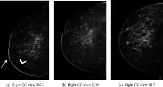
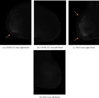
Female patient presented with a palpable lump in the right breast in early Nov 2016, followed by right breast erythematous skin discoloration and multiple dermal lesions spanning over 8cm.
Showing 13–24 of 36 cases