Case Feed
IsointenseTracer_849 · EXT-2026-328C84
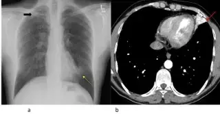
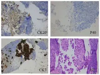
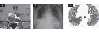
38-year-old male with mild exertional dyspnea.
IsointenseFocus_271 · EXT-2026-3D78C4
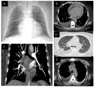
52 year-old man, heavy smoker (30 pack-years), diabetes, hyperlipidemia, obesity, presented with acute severe inspiratory dyspnea, chest pain, marked fatigability, and anxiety.
RadiantScan_702 · EXT-2026-2F484F
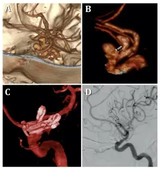
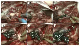
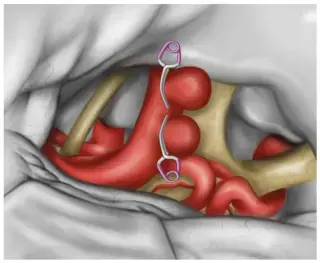
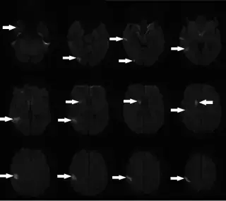
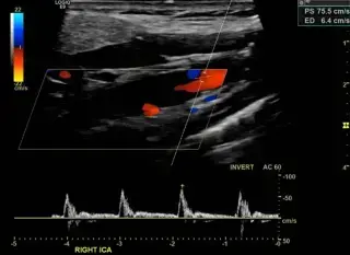
60-year-old female with a history of systemic arterial hypertension presented with an isolated transitory ischemic attack.
HyperdenseBeam_813 · EXT-2026-BF0FAE
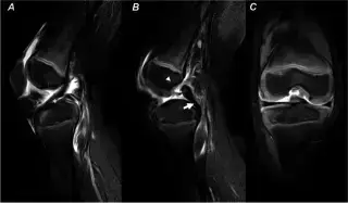
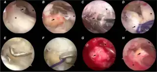
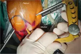
5-year-old girl with right knee pain and inability to bear weight after a hyperextension injury when her mother leaned on her while standing on one leg.
IsointenseFocus_271 · EXT-2026-293FE6
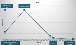
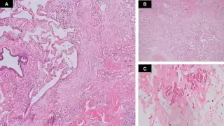
65-year-old man with headaches, diminished vision, loss of libido, erectile dysfunction since age 30. Diagnosed with sellar mass/hyperprolactinemia at age 44.
FocalMatrix_426 · EXT-2026-0489FB
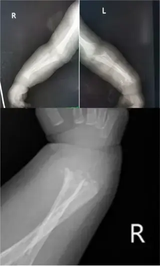
2-month old male infant presented for investigation of bilateral ulna and distal radius fractures. Also had noisy breathing, runny nose, reduced feeding. Mother noticed inability to move upper extremities and crying with passive movement a few days prior.
QuantumExplorer_803 · EXT-2026-955F1B
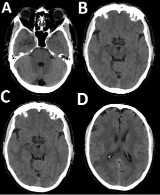
55-year-old male with sudden-onset left-sided facial droop, left arm paresthesia, uncoordinated gait, dysphasia, and sensory disturbances in the left arm. Family history of TIA.
ParenchymalViewer_870 · EXT-2026-D38E3A
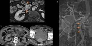
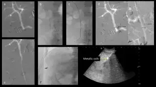
74-year-old female with history of total pancreatectomy for pancreatic head carcinoma, presented with ascites, diarrhoea, and lower limb oedema, leading to chemotherapy withdrawal.
AxiomNode_187 · EXT-2026-FBEE4C
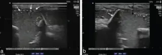
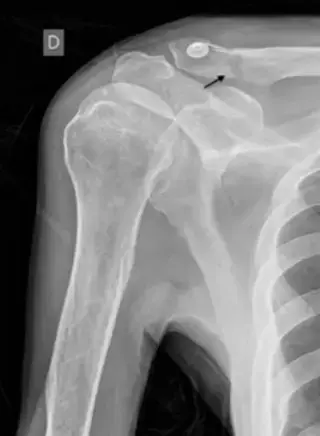
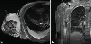
54-year-old male with fatigue and increasingly limited right arm mobility. History of fall 10 days prior, hitting head and right shoulder. Physical exam showed right facial hematoma, right shoulder contusion, and limited right shoulder motion.
RadiantScan_702 · EXT-2026-E31670
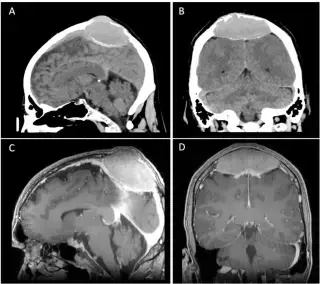
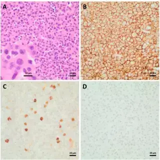
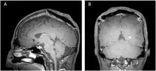
52-year-old male with a slowly progressive swelling of the skull, detected 10 months prior to consultation. Initially suspected benign lipoma by GP.
GantryGantry_464 · EXT-2026-711927
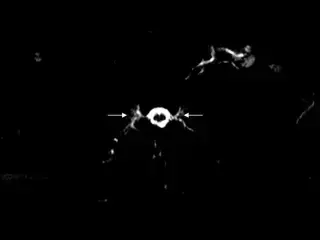
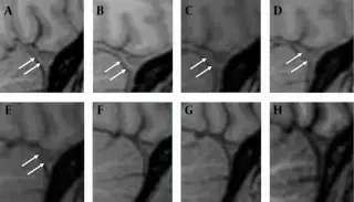
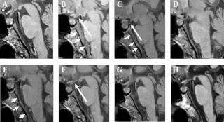
65-year-old man with 7 months of right fronto-parietal headache and neck soreness. Headache not posture-related, did not subside with analgesics. Developed orthostatic headache, SIH suspected.
HyperdenseBeam_813 · EXT-2026-ECDCD4
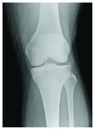
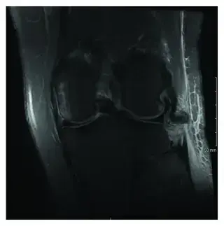
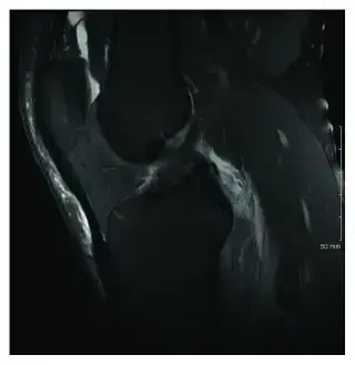
23-year-old male struck by motor vehicle, GCS 8. Left knee abrasions, lateral joint line tenderness with effusion. Right knee diffusely tender.
Showing 157–168 of 297 cases