Case Feed
HyperSlice_562 · EXT-2026-6F37AA
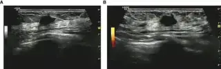
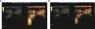
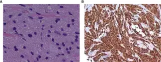
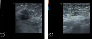
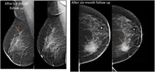
28-year-old woman, complaint: left breast lump for 1 month. Physical exam: 2x2 cm left breast mass, blurred borders, poor mobility, no tenderness.
ParenchymalSeeker_124 · EXT-2026-DC45E0
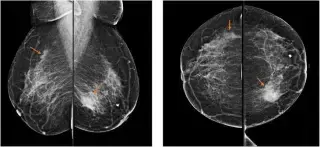
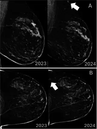
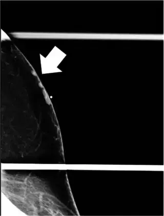
52-year-old female with a painful left breast mass for 5 days. No prior breast lumps, surgery, or family history of breast cancer.
ParenchymalSeeker_124 · EXT-2026-C7AD7E
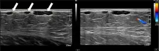
72-year-old woman, asymptomatic, annual screening mammogram revealed new mass.
ParenchymalFocus_598 · EXT-2026-52B3C0
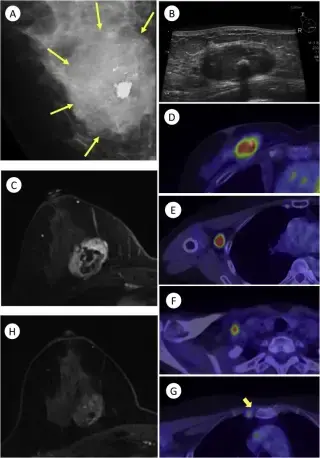
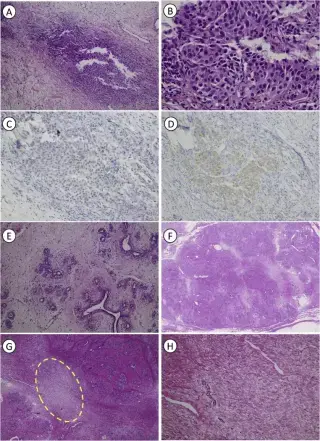
Patient with advanced triple negative breast cancer, multiple lymph node metastases, arising within a long-standing fibroadenoma.
HyperSlice_562 · EXT-2026-402800
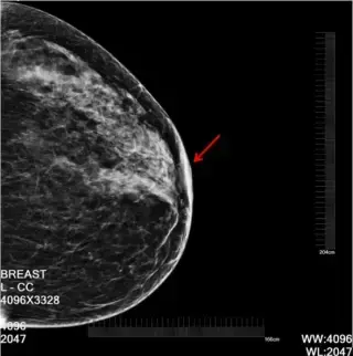
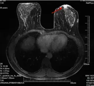
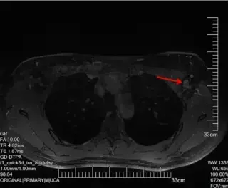
38F, recurrent left nipple fissure for 3 years, eczematous changes for 3 months. Noticed after lactation, mild discomfort, no discharge/bleeding. Topical meds no relief, led to nipple retraction.
HyperSlice_562 · EXT-2026-A274F4
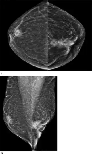
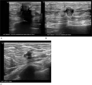
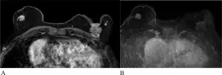
94-year-old male with bilateral palpable breast masses. History of bladder cancer, melanoma, squamous cell carcinoma. Negative for BRCA mutation. Bilateral breast masses and left axillary lymph nodes palpated. Bilateral inverted nipples, left nipple almost fully eroded.
ParenchymalFocus_598 · EXT-2026-AA8576
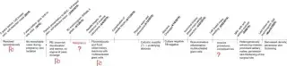
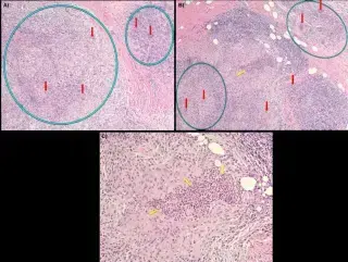
41-year-old female, G1P1 (C-section), with 3-month history of left breast pain, swelling, chills, night sweats, skin changes, and self-detected mass. Previous similar episode 5 years prior.
TeslaWave_474 · EXT-2026-74EA0B
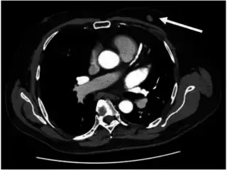
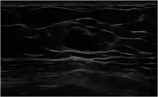
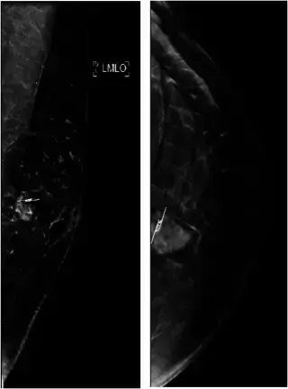
63-year-old man referred for an incidental CT finding of an enlarging left anterior chest wall nodule. History of distal pancreatectomy/splenectomy for pancreatic neuroendocrine tumour 12 years ago, and insulin-dependent diabetes mellitus.
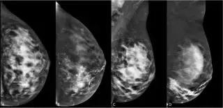
ParenchymalFocus_598 · EXT-2026-32DF64
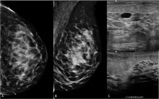
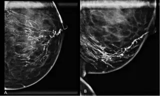
37-year-old female, 22 weeks pregnant, presented with 2 months of spontaneous bloody left nipple discharge. No breast pain, lump, or skin changes. No breast cancer history.
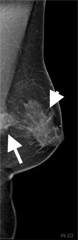
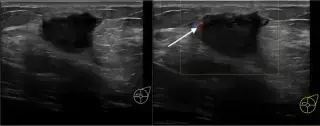
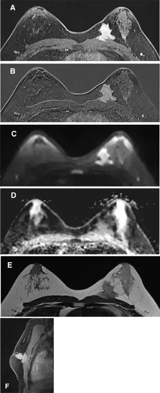
HyperSlice_562 · EXT-2026-F16F71
40-year-old man with a 4-month history of self-palpated left breast mass. Known chronic bilateral gynecomastia and insulin-treated diabetes. No personal/family breast cancer history.
SpecularPhantom_904 · EXT-2026-982994
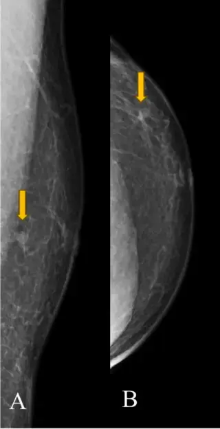
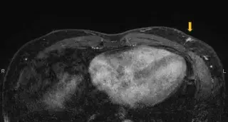
62-year-old male presented with a lump in the lower outer quadrant of his breast. Family history significant for three relatives with breast cancer.
ParenchymalSeeker_124 · EXT-2026-EEE8C7
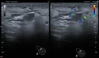
32-year-old lactating female with 6-month history of gradually increasing right breast swelling, 1 month of rapid increase, no pain or bleeding. No comorbidities or family history of breast cancer.
Showing 1–12 of 36 cases