Case Feed
EchoScout_273 · EXT-2026-59A5C5
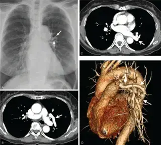
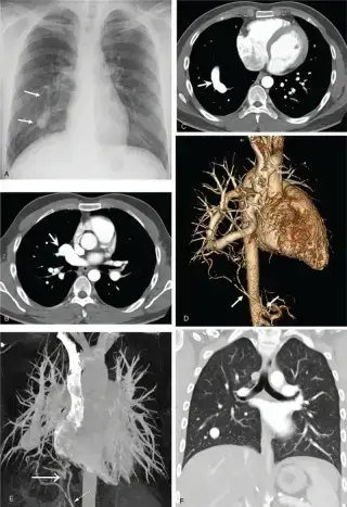
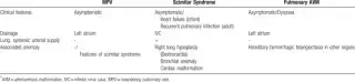
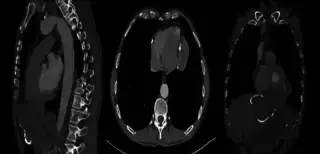
71-year-old woman, abnormal chest radiographic finding on medical check-up. Previously healthy, no specific medical history or respiratory symptoms.

GantryWindow_627 · EXT-2026-BD61D8
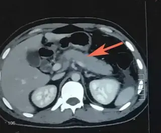
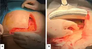
Male, presented with 1-day history of sudden onset, sharp, severe epigastric and left upper quadrant pain radiating to the back, aggravated by meals, associated with non-projectile, non-bilious vomiting.
EchoScout_273 · EXT-2026-614695
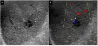
65-year-old male with two months of right upper quadrant pain, left-sided chest pain, and epigastric discomfort. No past medical history. Physical exam showed epigastric dullness. Labs normal.
ParenchymalFocus_598 · EXT-2026-63671B
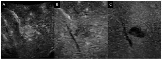
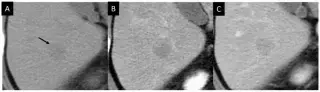
An 84-year-old man, asymptomatic, underwent abdominal ultrasound for diabetes mellitus screening. History of diabetes mellitus and hypertension. No alcohol, blood transfusions, or remarkable family history.
HounsfieldMatrix_926 · EXT-2026-D2011B
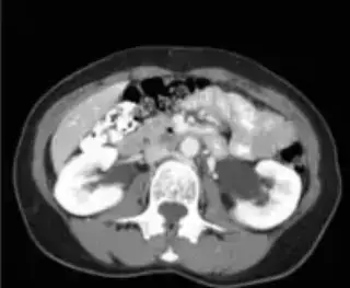
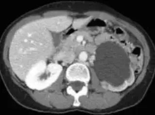
62-year-old woman with brief left flank pain. No prior urologic history of colic, urolithiasis, or infection. Serum creatinine 1.0 mg/dl, urine culture negative.
GantryWindow_627 · EXT-2026-A13227
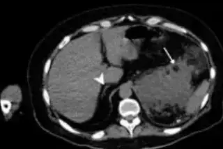
47-year-old male with hypertension refractory to several drugs.
GantryWindow_627 · EXT-2026-D72D61
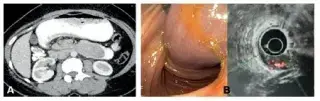
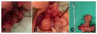
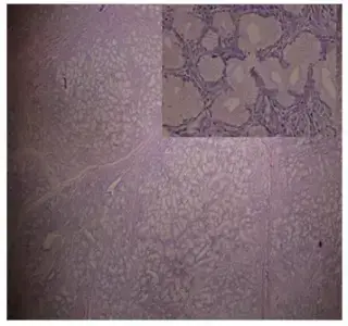
33 year old female with vague epigastric discomfort, weakness, breathlessness on exertion, 3-4 episodes of melena, and intermittent non-bilious vomiting for 45 days. Exam showed pallor, mild epigastric fullness. Hb 6.7 g/dl.
QuantumNode_728 · EXT-2026-2ECEDC
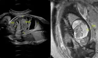
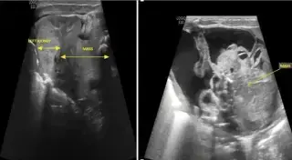
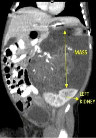
Female neonate, delivered at 39 weeks. Fetal USG at 31 weeks showed a left abdominal cystic structure. Presented with abdominal mass & mild abdominal distension.
ParenchymalViewer_870 · EXT-2026-10A81A
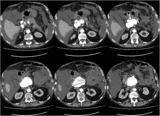
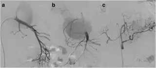
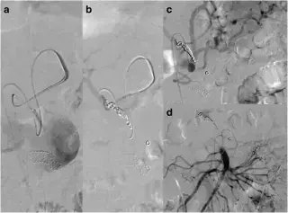
67-year-old man with diffuse abdominal pain, vomiting, diarrhea, and 4 kg weight loss over weeks. History of alcohol abuse.
SpectralScan_327 · EXT-2026-1E8FE6
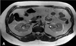
62-year-old man with hypertension and obesity developed persistent gross hematuria after a bicycle fall.