Case Feed
ParenchymalFocus_598 · EXT-2026-F1FD01
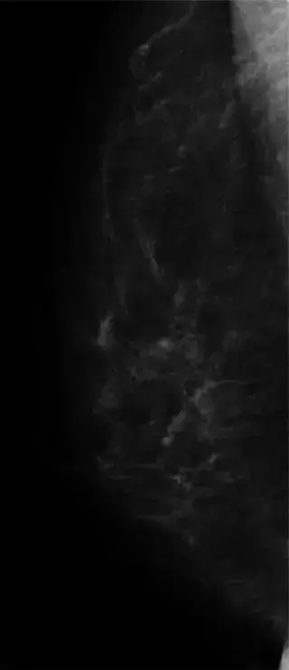
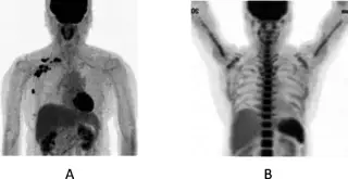
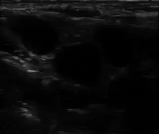
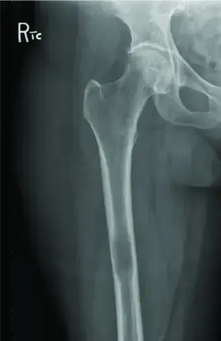
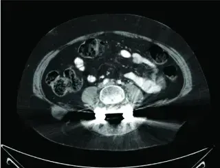
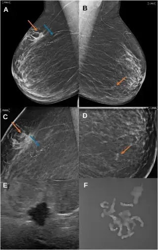
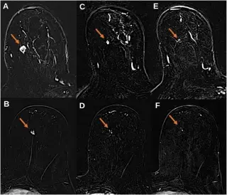
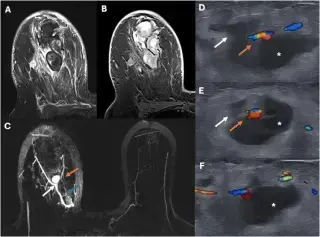
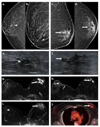
62F with history of left micro invasive breast cancer 15 years prior, treated with breast-conserving therapy, sentinel node biopsy, and 5 years adjuvant tamoxifen. Annual check-ups.
ParenchymalFocus_598 · EXT-2026-594E7D
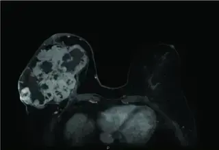
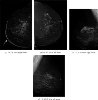
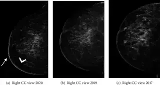
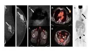
58 year-old woman with no prior mammograms presented with increasing pain in her right breast for 7 months. Physical exam showed enlarged breast with multiple visible nodules.
ParenchymalFocus_598 · EXT-2026-978CA4
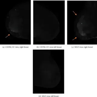
Female patient presented with a palpable lump in the right breast in early Nov 2016, followed by right breast erythematous skin discoloration and multiple dermal lesions spanning over 8cm.
HyperSlice_562 · EXT-2026-F16F71
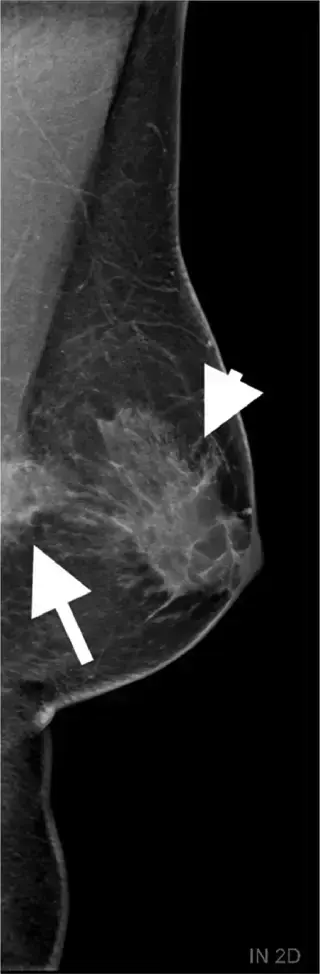
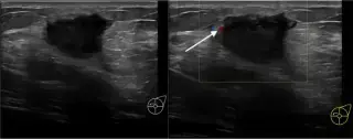
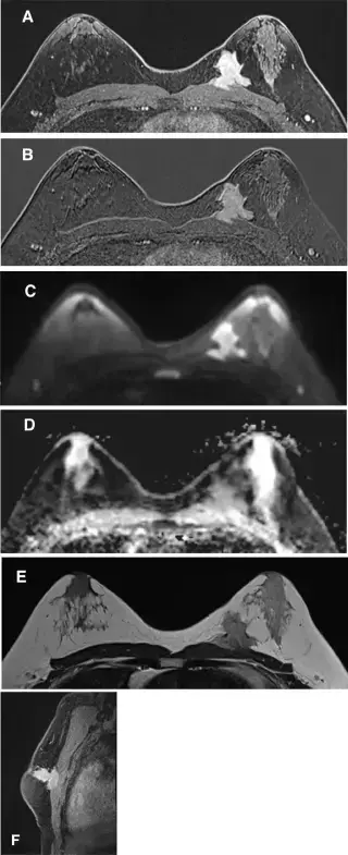
40-year-old man with a 4-month history of self-palpated left breast mass. Known chronic bilateral gynecomastia and insulin-treated diabetes. No personal/family breast cancer history.
HyperSlice_562 · EXT-2026-279D0C
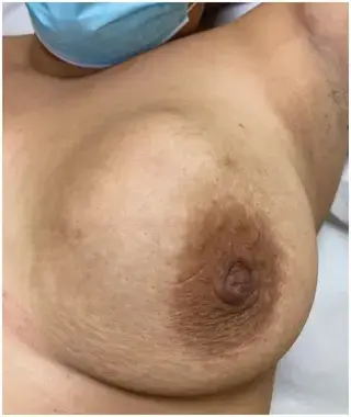
64F, postmenopausal, hormonal substitution, hx surgically treated VIN 3, family hx gynecological cancers, positive breast cancer screening.
HyperSlice_562 · EXT-2026-ABC594
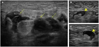
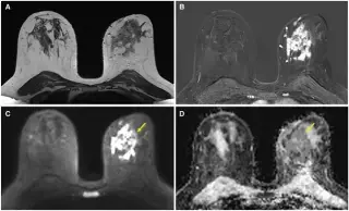
24-year-old female smoker, presented with a suddenly appearing indurated palpable mass in the upper-inner quadrant of her left breast with associated mild erythema, 48h after COVID-19 vaccination.
ParenchymalFocus_598 · EXT-2026-8F92C0
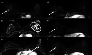
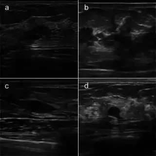
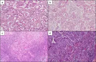
A 26-year-old woman with a BRCA1 pathogenic variant undergoing annual breast screening.
ParenchymalFocus_598 · EXT-2026-9165A3
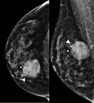
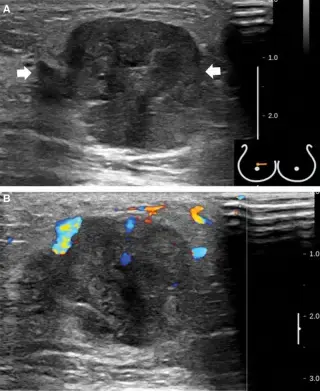
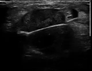
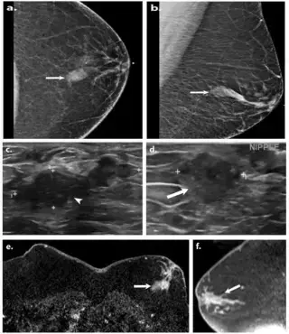
47F, premenopausal, with a painless palpable right breast mass for 7 months. No significant medical/family history of breast cancer. No skin changes, nipple discharge, or palpable axillary nodes.
TeslaWave_474 · EXT-2026-BFE948
61-year-old male with hypertension and type 2 diabetes mellitus, presenting with a one-week history of a non-tender palpable left breast mass.
HyperSlice_562 · EXT-2026-ADCCEB
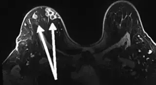
50-year-old female with multiple palpable nodules of the right breast. History of hypertension, hyperlipidemia, diabetes, tetralogy of Fallot with bioprosthetic valve, and bilateral silicone breast implants.
ParenchymalSeeker_124 · EXT-2026-8D54AE
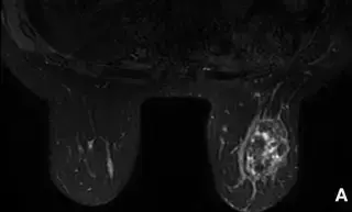
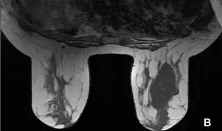
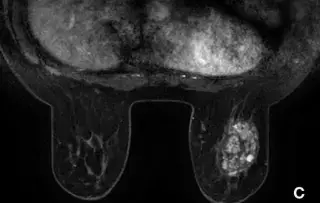
50-year-old woman with a palpable left breast mass and radiating left arm pain for months. History includes no previous pregnancy, no family pathology, tubal ligation, and total hysterectomy for fibroids.
HyperSlice_562 · EXT-2026-4A656D
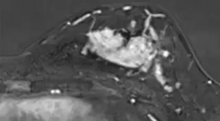
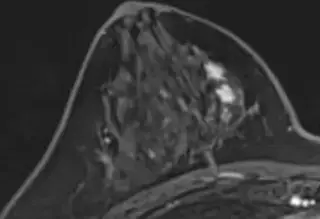
36-year-old woman with a palpable mass in the left breast.