Key Images. Click to enlarge
Clinical History
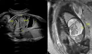
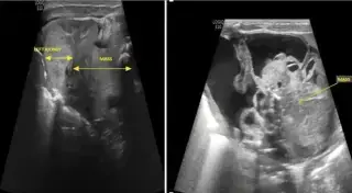
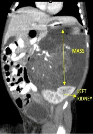
Female neonate, delivered at 39 weeks. Fetal USG at 31 weeks showed a left abdominal cystic structure. Presented with abdominal mass & mild abdominal distension.
Write your interpretation
Sign in to submit your read. The original report is revealed only after you submit.